Welcome to the Amira-Avizo Software Use Case Gallery
Below you will find a collection of use cases of our 3D data visualization and analysis software. These use cases include scientific publications, articles, papers, posters, presentations or even videos that show how Amira-Avizo Software is used to address various scientific and industrial research topics.
Use the Domain selector to filter by main application area, and use the Search box to enter keywords related to specific topics you are interested in.
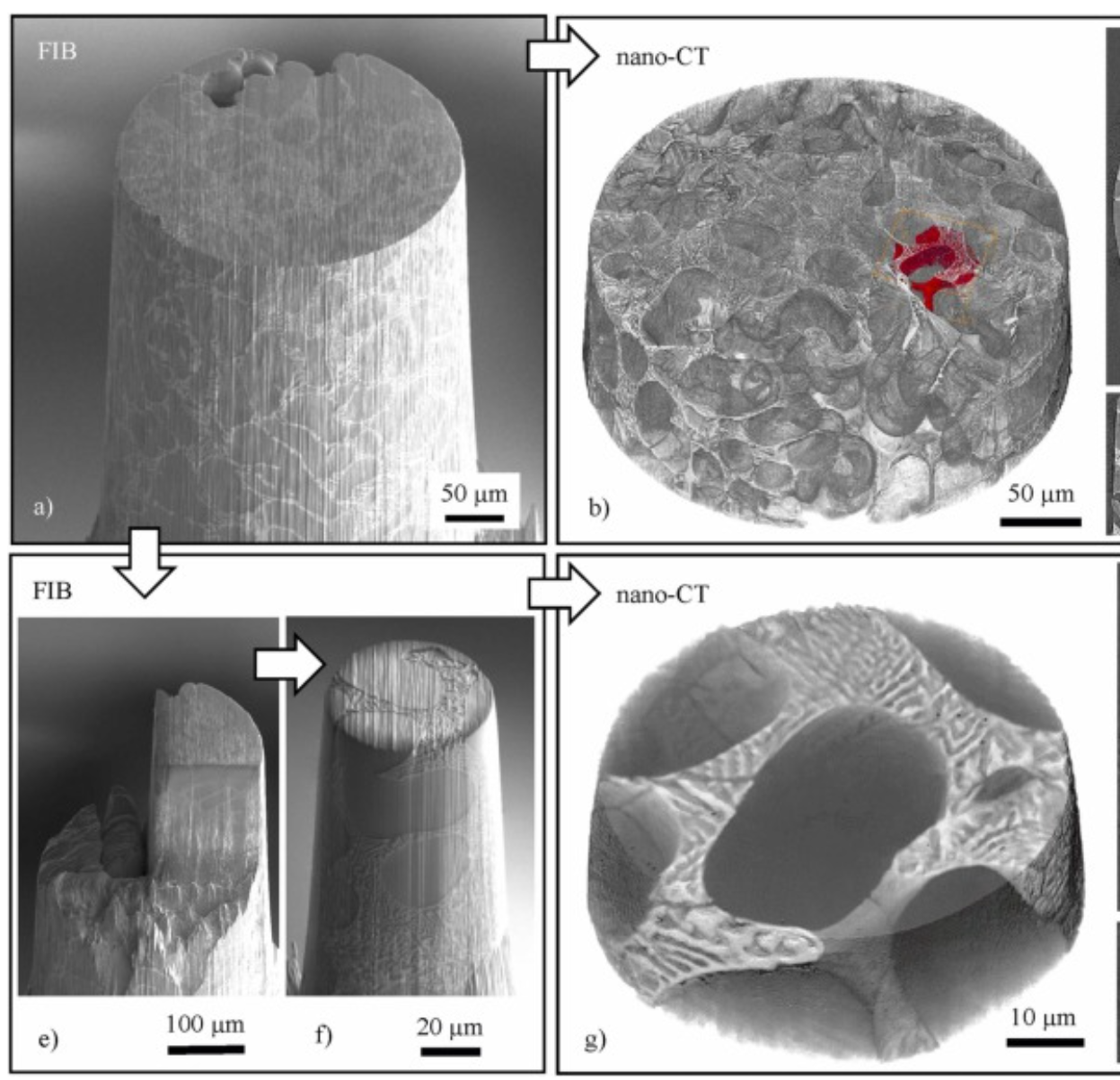
Three-dimensional imaging of microstructural evolution in SEM-based nano-CT
Scanning electron microscopy (SEM) is a powerful and versatile technique for materials characterization and present in many laboratories. The integration of an X-ray target holder and detector allows expanding the modalities of SEM by X-ray imaging. These little hardware adaptations enable radiography ... Read more
Jonas Fell, Christoph Pauly, Michael Maisl, Simon Zabler, Frank Mücklich, Hans-Georg Herrmann
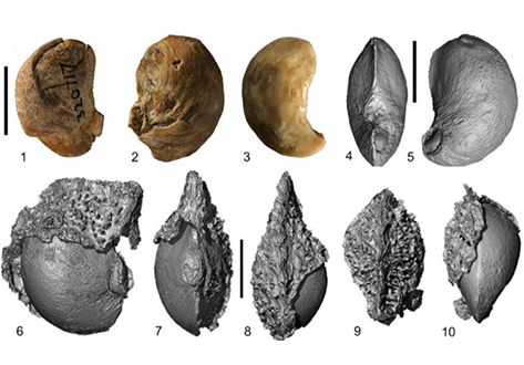
Fossil fruits formerly described as cashews from the Oligocene of Peru are reinvestigated based on the original specimens and newly collected materials. The recovery of an outer spiny layer, preserved in the sedimentary molds surrounding the locule casts, indicates that these disseminules do not represent Anacardium. Imagery from nano-CT scans of the specimens documents a distinctive morphology which does not resemble any fruits or seeds of Anacardiaceae. We describe the morphology in more de... Read more
STEVEN R. MANCHESTER, BEHNAZ BALMAKI
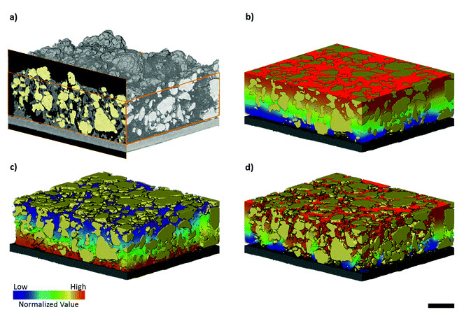
The microstructural degradation of a composite silicon electrode at different stages in its cycle life was investigated in 3D using X-ray nano-computed tomography. A reconstructed volume of 36 μm × 27 μm × 26 μm from the composite electrode was imaged in its pristine state and after 1, 10 and 100 cycles. Particle fracturing and phase transformation was observed within the electrode with increased cycling. In addition, a distinct, lower X-ray attenuating phase was clearly resolved,... Read more
Oluwadamilola O. Taiwo, Melanie Loveridge, Shane D.Beattie, Donal P.Finegan, Rohit Bhagat, Daniel J.L.Brett, Paul R.Shearing
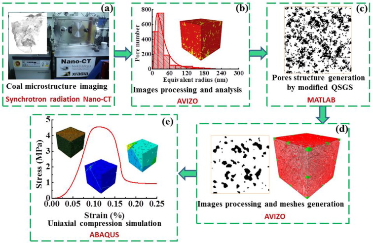
Impact of nanopore structure on coal strength
In China’s energy consumption structure, coal is the main energy source, accounting for about 60% of primary energy consumption. Coal is a porous medium with complex pore structures. Nanopore structure in coal particle is the basic underlying factor driving coal particle strength. A better knowledge of nanopore structure – coal particle strength correlation is of great significance for coal mining and other fields of engineering problems.
Read more
Yixin Zhao - Liang Yuan - QuanXue
Three-dimensional image based modelling of transport parameters in lithium–sulfur batteries
An elemental sulfur electrode was imaged with X-ray micro and nano computed tomography and segmented into its constituent phases. Morphological parameters including phase fractions and pore and particle size distributions were calculated directly from labelled image data, and flux based simulations were performed to determine the effective molecular diffusivity of the pore phase and electrical conductivity of the conductive carbon and binder phase, D... Read more
Chun Tan, Matthew D. R. Kok , Sohrab R. Daemi , Daniel J. L. Brett and Paul R. Shearing
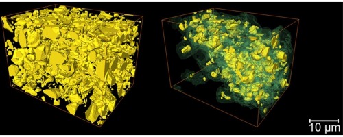
Visualizing the Carbon Binder Phase of Battery Electrodes in Three Dimensions
This study presents a technique to directly characterize the carbon and binder domain (CBD) in lithium-ion (Li-ion) battery electrodes in three dimensions and use it to determine the effective transport properties of a LiNi0.33Mn0.33Co0.33O2 (NMC) electrode. X-ray nanocomputed tomography (nano-CT) is used to image an electrode composed solely of carbon and binder, whereas focused ion beam–scanning electron microscopy is used to analyze cross-sect... Read more
Sohrab R. Daemi, Chun Tan, Tobias Volkenandt, Samuel J. Cooper, Anna Palacios-Padros, James Cookson, Dan J. L. Brett, and Paul R. Shearing
Micron-scale crack propagation in laser-irradiated enamel and dentine studied with nano-CT
The aim of this study was to see the effect of Er:YAG laser irradiation in dentine and compare this with its effect in enamel. The mechanism of crack propagation in dentine was emphasised and its clinical implications were discussed. A possible mechanism is that laser radiation is transmitted down the dentinal tubules causing micro-cracks to form in the dentinal tubule walls that tend to be limited to this region. Crack might be a source of fracture as it represents a weak point and subsequen... Read more
Abtesam Aljdaimi, Hugh Devlin, Mark Dickinson, Timothy Burnett, Thomas J. A. Slater
Ptychographic X-ray computed tomography (PXCT) is a quantitative imaging modality that non-destructively maps the 3D electron density inside an object with tens of nanometers spatial resolution. This method provides unique access to the morphology and structure of the osteocyte lacuno-canalicular network (LCN) and nanoscale density of the tissue in the vicinity of an osteocyte lacuna. Our findings indicate that PXCT can non-destructively provide detailed, nanoscale information on the 3D org... Read more
Antonia Ciani, Hechmi Toumi, Stéphane Pallu, Esther H.R.Tsai, Ana Diaz, Manuel Guizar-Sicairos, Mirko Holler, Eric Lespessailles, Cameron M.Kewish | Synchrotron Soleil, France